I always thought that bipedalism was the boring part of human evolution, a bit of replumbing that had to happen before we could invent language and control fire. But then I realized that the transition to bipedalism was still causing health problems in humans, and the subject got a lot more interesting.
To be sure, the move to bipedalism was an extraordinary development, unprecedented among mammals. It allowed us to carry baskets and spears in one direction, and tubers and game in the other. But our ape-like ancestors accomplished it by modifying a body plan that was previously arboreal. Our feet, legs, hips, and backs were all MacGyvered, to varying degrees of success.
Biological anthropologists noticed these shortcomings and their causes a while back. In a 1951 Scientific American article, Wilton M. Krogman pointed out many of the defects that arise from quirks in our anatomy, including hernias, sacroiliac joint pain, hemorrhoids, varicose veins, fallen arches, and a whole host of back troubles (Krogman 1951).
Starting off on the Right Foot
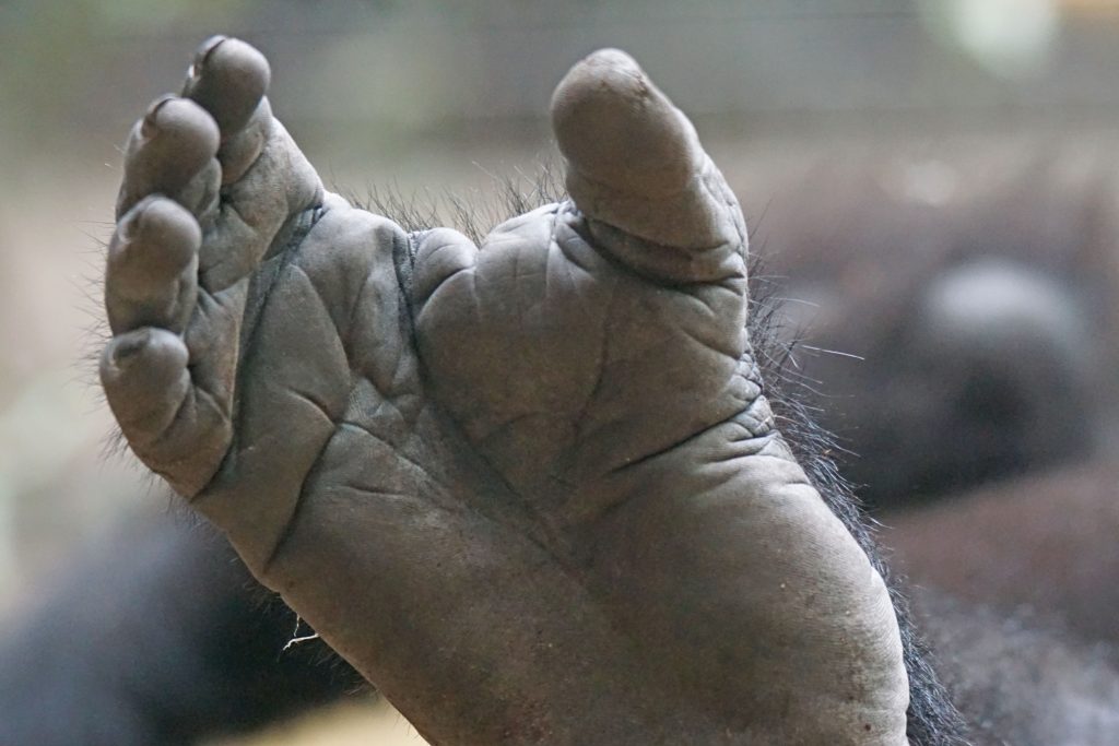
Since primates appeared on the evolutionary scene, their defining characteristic has been tree-climbing. But humans keep their feet firmly on the ground (usually), and the design of our feet has changed accordingly.
Unlike our primate cousins, we have big toes that line up nicely with our other toes, and our feet are useless for grasping branches. We have arches to help us dissipate energy, and spring forward when we walk. And our calcaneus (heel bone) is proportionally larger in humans than in any other mammal. Biological anthropologist Bruce Latimer pointed out that, “[A] 350 pound male gorilla has a calcaneus with a smaller cross section than does a human female weighing 100 pounds (Latimer 2005).”
But just because our feet have been sensibly altered doesn’t mean they’re perfectly suitable. For starters, we still have a lot of bones – and a lot of joints – in an appendage that would ideally be stiff and springy. As DeSilva pointed out, “If you were going to start from scratch, there’s no way you’d make a foot out of 26 parts. And if you did, you wouldn’t be accused of being intelligent (Boston University 2019).”
It’s tempting to think that our bunions, heel spurs, and fallen arches are the consequence of wearing loafers and walking across concrete sidewalks, but the fossil record suggests that many modern foot problems predate our species. Two ancient hominin fossils have evidence of ankle breaks, one had osteoarthritis in the foot, and one had a compression fracture (Boston University 2019). Considering we only have a small fossil sample to go on, the rates of injury are particularly high.
I’ve noted that plantar fasciitis is often an unfortunate consequence of a sitting disability. Apparently, it’s also an unfortunate consequence of an upright life. Latimer notes that, “Other maladies that occur only in humans are plantar fasciitis and the development of ‘heel spurs.’ Both of these conditions result from repeated tension on the plantar aponeurosis, a strong, inelastic ligamentous structure that stretches from the plantar margin of the calcaneus to the bases of the proximal phalanges (Latimer 2005).”
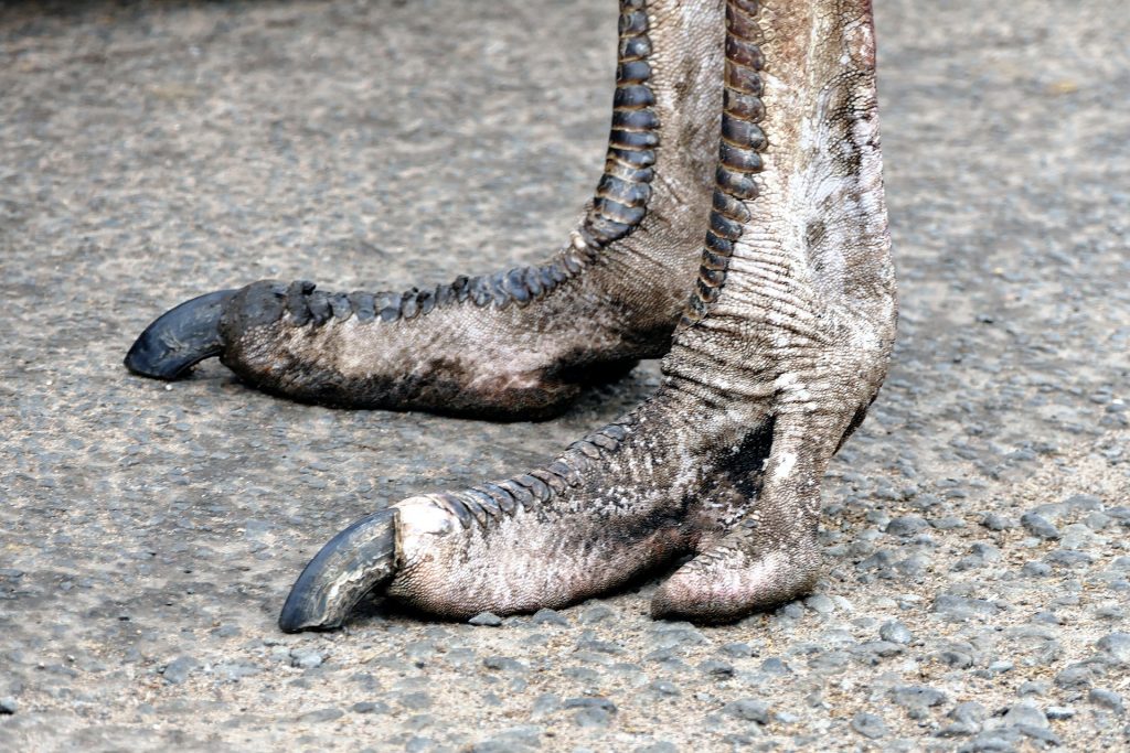
There are other design options for bipedal feet that are less prone to these sorts of injuries. Just ask the ostrich. They run quite efficiently on two toes, and they’ve moved their ankle joints halfway up their legs.
Our Achilles’ Heel
For many athletes, their Achilles’ heel is…well, their Achilles’ heel. In apes, the Achilles’ tendon is short, or missing altogether. But for bipedal humans, it’s the strongest tendon in the body. While we’re walking, it acts as a spring to help propel us forward (Malvankar and Khan 2011).
It is also, unfortunately, prone to injury. In an ironic twist, active people (who have thicker Achilles tendons on average) are more likely to be injured than couch potatoes. And unfortunately, the Achilles tendon is not that good at repairing itself. Some sections don’t have much of a blood supply, and damaged tissues are repaired using a different type of collagen that’s less good at stretching.
For top athletes, Achilles tendon injuries can be serious. A study of injured NFL players showed that 36% never played professionally again, and those who did return never played as well as they had before (Malvankar and Khan 2011).
A study of 18 Irish dancers showed that 14 of them had Achilles tendinopathy (even though 7 of them reported no pain). Seven of the dancers also had plantar fasciitis, so this study confirmed the commonsense conclusion that Irish dancing is tough on your feet (Walls, et al. 2010).
Part of the problem is that we place so much pressure on our feet and legs. Quadrupedal animals use the muscles in their shoulders and backs to absorb and dissipate the shocks from each step (Latimer 2005). But humans can’t do this. When we walk, we hit the ground one foot at a time. We have a few shock absorbing structures, like our quadriceps muscles and the curve in our spine, but we’re still prone to injury in our feet, knees, and hips.
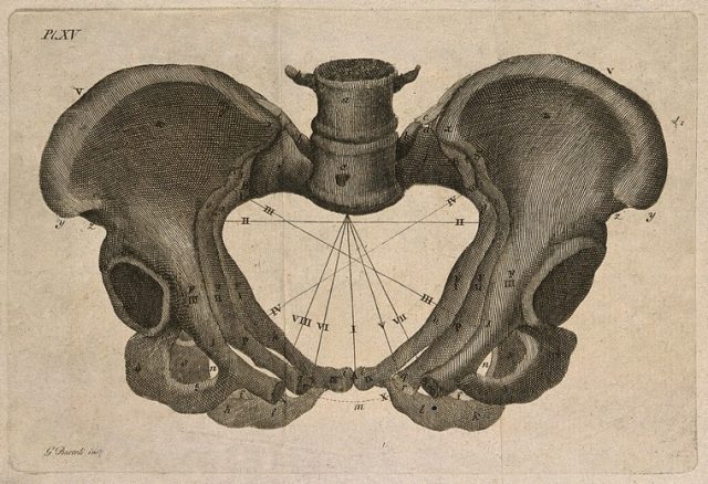
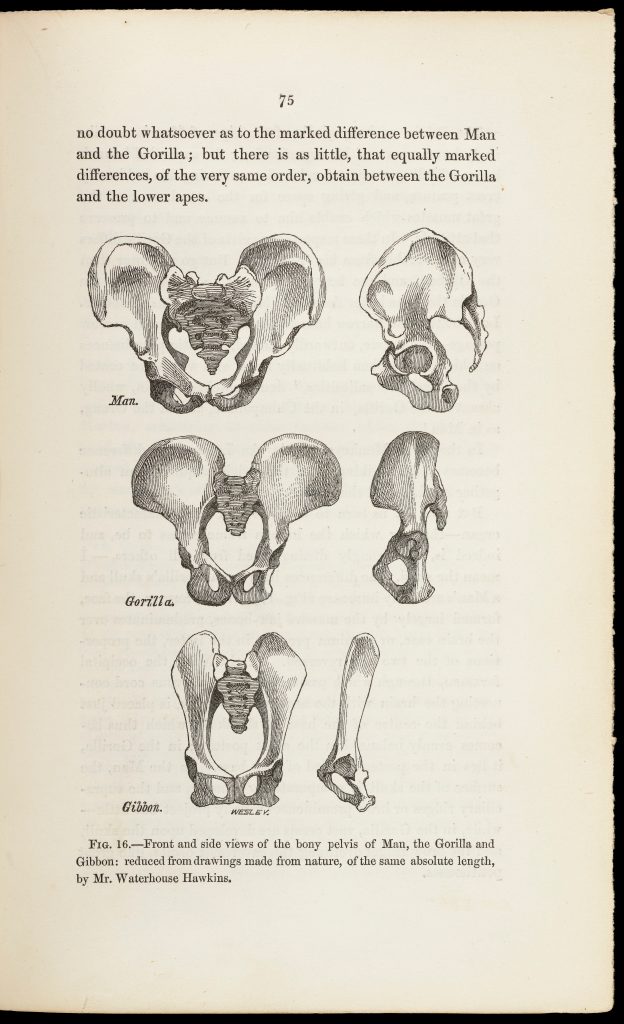
The Pelvis
Our pelvis is one giant compromise. It’s shorter and broader than a chimpanzee or gorilla pelvis, which allows for better energy transfer when we walk. In humans, the pelvis also holds up our internal organs, provides outlets for our urinary and digestive tracts, serves as an attachment site for our leg muscles, supports our upper body weight, and gives us something to sit on. And, in women, it lets a baby out (Krogman 1951).
In his delightfully engaging book, The Body: A Guide for Occupants, Bill Bryson nicely summarizes the challenge:
The problem with human childbirth is cephalopelvic disproportion. In simple terms, a baby’s head is too big for smooth passage through the birth canal, as any mother will freely attest. The average woman’s birth canal is about an inch narrower than the width of the average newborn’s head, making it the most painful inch in nature. To squeeze through this constricted space, the baby much execute an almost absurdly challenging ninety-degree turn as it proceeds through the pelvis. If ever there was an event that challenges the concept of intelligent design, it is the act of childbirth. No woman, however devout, has ever in childbirth said, “Thank you, Lord, for thinking this through for me (Bryson 2019).”
The ability to reproduce is non-negotiable for evolutionary success. Yet, humans are amazingly bad at it. We give birth to giant-headed babies that require years of constant care. Newborn gorilla babies are, on average, 2.7% as big as their mother. Newborn chimpanzees are 3.3% as big. Newborn humans are 6.1% as big as their mothers, and completely helpless (Boston University 2019).
The size of the baby compared to the size of its mother’s pelvis means that childbirth is uniquely dangerous for humans. Even today, in countries with the highest maternal mortality rates, over 1% of women die in childbirth (Unicef 2019). And that’s with modern medicine (however unevenly available).
Historical rates are harder to calculate, so we don’t know how many Cro-Magnon mothers perished along with their babies. But there are a few useful estimates to provide a comparison. In England in the first half of the 18th century, maternal mortality rates were estimated to be 1,050 per 100,000 live births (McFadden and Oxenham 2019). Estimates in modern hunter-gatherers range from 670 deaths for the Ache of Paraguay, to 1,022 in the Hadza of Tanzania (Curnoe 2016).
A Fractured Present
Even osteoporosis may be a weird quirk of evolution. Many human bones, especially those in the foot, ankle, and knee, are larger, but less dense, than their ape counterparts. The added surface area in human bones makes it easier for minerals to be leached out and accelerates bone loss (Latimer 2005).
One small study compared human vertebrae with those of gibbons, orangutans, chimpanzees, and gorillas. It found that humans had larger vertebrae for their body mass, but the human vertebrae were less dense, and had a thinner layer of tough cortical bone on the outside. This could possibly be why humans are prone to vertebral fractures.
The spongy, trabecular bone that makes up most of our vertebrae is better at absorbing shocks, because it can deform and spring back. Cortical bone is stiffer, but it’s also more likely to shatter if the stress is too great.
The authors noted that other apes do get osteoporosis, sometimes severely, yet scientists haven’t found a spontaneous vertebral fracture in either captive or wild apes (Cotter, et al. 2011).
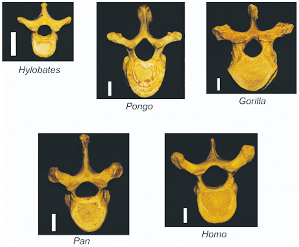
Photo from “Human Evolution and Osteoporosis-Related Spinal Fractures.”
What are the Practical Applications?
I watched one panel on biological anthropology where an audience member asked, “How do these evolutionary legacies have clinical applications?” The panelists couldn’t come up with a compelling answer. Neither could I. As a patient, I felt that concepts in evolutionary medicine were useful, but I couldn’t think of any definite reason why this should be.
I eventually realized that knowing my design flaws helps to put injuries in context. Injuries and illnesses can feel like an act of fate, and in some sense, they are. But before we were born, primates spent millions of years evolving into humans. When we arrive at a moment where a bone breaks or a foot hurts, it’s like we’ve arrived onstage during the last five minutes of a play. The setup has already happened, we just need to deliver the punchline.
Of course, I have never asked a woman in childbirth whether she feels the same way.
References
Boston University. 2019. Boston University Dialogues in Biological Anthropology Number 6 – Part 1. April 2. Accessed January 6, 2021. https://www.youtube.com/watch?v=QKL061Rnk-I.
Bryson, Bill. 2019. The Body: A Guide for Occupants. Knopf Doubleday Publishing Group. https://www.penguinrandomhouse.com/books/239783/the-body-by-bill-bryson/.
Cotter, Meghan M, David A Loomis, Scott W Simpson, Bruce Latimer, and Christopher J. Hernandez. 2011. “Human Evolution and Osteoporosis-Related Spinal Fractures.” PLoS ONE. doi:10.1371/journal.pone.0026658.
Curnoe, Darren. 2016. Need help with that delivery? Call the monkey midwife! May 2. Accessed January 7, 2021. https://theconversation.com/need-help-with-that-delivery-call-the-monkey-midwife-58630.
Gibbons, Ann. 2013. “Human Evolution: Gain Came With Pain.” Science, February 16. Accessed January 5, 2021. https://www.sciencemag.org/news/2013/02/human-evolution-gain-came-pain.
Krogman, Wilton M. 1951. “The Scars of Human Evolution.” Scientific American 185 (6): 54-57. https://www.scientificamerican.com/article/the-scars-of-human-evolution/.
Latimer, Bruce. 2005. “The Perils of Being Bipedal.” Annals of Biomedical Engineering 33 (1): 3–6. doi:10.1007/s10439-005-8957-8.
Malvankar, S, and W S Khan. 2011. “Evolution of the Achilles tendon: The athlete’s Achilles heel?” The Foot (Elsevier) 21 (4): 193–197. doi:10.1016/j.foot.2011.08.004.
McFadden, Clare, and Marc F Oxenham. 2019. “The Paleodemographic Measure of Maternal Mortality and a Multifaceted Approach to Maternal Health.” Current Anthropology 60 (1). doi:10.1086/701476.
Unicef. 2019. Maternal Mortality. September. Accessed January 6, 2021. https://data.unicef.org/topic/maternal-health/maternal-mortality/.
Walls, R J, S A Brennan, P Hodnett, J M O’Byrne, S J Eustace, and M M Stephens. 2010. “Overuse ankle injuries in professional Irish dancers.” Foot and Ankle Surgery (Elsevier) 16 (1): 45–49. doi:10.1016/j.fas.2009.05.003.
One thought on “Bipedalism: The Downsides of an Upright Life”