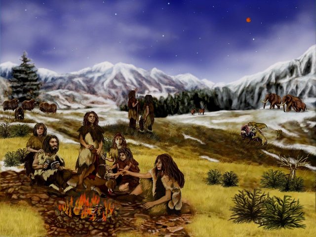
Over the Christmas vacation, I got sucked into reading about the devastated spines of our extinct hominin relatives. Partly, this was because I wanted to know whether back problems truly are a modern problem, but I’m also fascinated by any subject that starts with “paleo-“ and ends with “-ology.” Paleopathology? I didn’t know that was a thing, but when I found out, I spent weeks spelunking in that rabbit hole.
As it turns out, there were a lot of things wrong with ancient spines. A pristine spine is the exception, not the norm. Martin Haeusler, an evolutionary morphology scholar whose work I relied on heavily for this article, noted that only three specimens with decent spinal remains lack any noteworthy pathologies (Haeusler, Spinal Pathologies in Fossil Hominins 2019).
This article is devoted to the bad spines of hominins that walked upright, but weren’t fully human in the sense of being Homo sapiens. That may sound like an oddly specific topic, but it still breezes through about 3.5 million years of history.
I was impressed that scientists know as much as they do about the spinal health of prehistoric individuals. Vertebrae are full of spongy, trabecular bone that doesn’t fossilize as well as other hard parts, like teeth or thigh bones. They also have an arch with a few bony promontories, which are likely to break off, get crushed, or otherwise disintegrate. Scientists are rarely blessed enough to find an entire spine, so they have to do their best with what they find.
Individual Attention
One aspect of paleopathology that I very much enjoyed was the scrutiny that was given to each fossil. They were excavated, reassembled, measured, photographed, scanned, cast, and modeled using computer software.
This is completely different than the approach to modern-day back problems. When I see articles about clinical trials, patients get collapsed into broad categories based on symptoms, and occasionally pathologies. “Sciatica,” “herniated disc,” And the annoyingly vague, “low back pain.” There’s rarely any space devoted to analyzing individual differences.
As one author put it, “A key challenge in mapping the progress of evolution is the limited number of specimens and the poor condition of most samples. Unlike medical research where statistical significance is critical for conclusions, the foundation of evolution research is the development of theories or hypotheses based on scant physical artifacts (Sparrey, et al. 2014).”
The paleo approach, with attention given to each individual specimen, even to each individual vertebra, feels more natural to me as a patient. I don’t spend much time thinking about what’s statistically significant across a population. I spend a lot of time thinking about the quirks in my own spine. And while I haven’t conducted a survey, I’d wager that most back pain patients feel the same way.
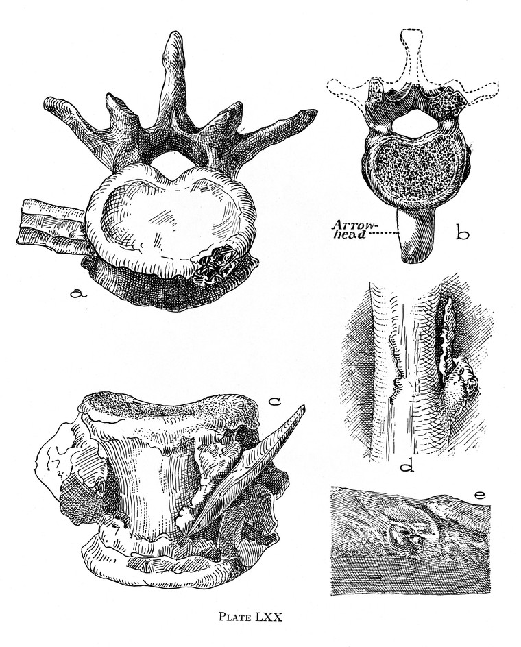
Photo courtesy of the Wellcome Collection.
Lucy the Hunchback?
The earliest noteworthy fossil vertebrae come from a 3.6 million-year-old Australopithecus afarensis specimen. However, only the C2–C6 vertebrae were preserved, and most of the bony protrusions had worn away (Ryan and Sukhdeo 2015). From this, scientists noticed that the pattern of osteophyte (sometimes called “bone spur”) growth more closely resembled modern humans than apes, which supported the notion that australopithecines walked in a human-like fashion (Haeusler, Spinal Pathologies in Fossil Hominins 2019).
The famous Lucy, another member of Australopithecus afarensis, died about 3.2 million years ago while in her mid-20s. She left us nine vertebrae, though scientists don’t agree on which ones. Most of the vertebrae were between T6–T10, along with 1 or 2 lumbar vertebrae (Meyer, et al. 2018).
Lucy might have had a hump-backed appearance. Her thoracic vertebrae were wedged so much that researchers suggested she had Scheuermann’s disease (Haeusler, Spinal Pathologies in Fossil Hominins 2019), which is characterized by excessive curvature of the spine in the upper torso. But like much else with the mysterious Lucy, this is still up for debate.
By the way, Scheuermann’s disease might be the most common spinal condition I’d never heard of. Today, it’s estimated to affect between 4–8% of adolescents. I couldn’t find good figures for adults, since it usually starts (and is most treatable) in young people. There hasn’t been much research done on the long-term effects (especially when people decide to live with it, rather than asking their doctors for help), but it appears that the problems are usually cosmetic, rather than functional (Tsirikos and Jain 2011).
Photo courtesy of MusicNewz via Wikimedia Commons.
Scheuermann’s disease is also a common diagnosis in the bipedal hominin fossil record. At least three other isolated vertebrae from different Australopithecus afarensis individuals also show some characteristics of the disease, which makes you wonder whether Lucy and her friends would have classified it as such.
One juvenile specimen of Australopithecus africanus from South Africa also met the full criteria for Scheuermann’s disease.
The Prehistory of Herniated Discs
I have a personal interest in herniated discs, but I didn’t expect to find much evidence of them in the fossil record. After all, the soft tissues of intervertebral discs don’t lend themselves to preservation. But here, scientists surprised me by their cleverness. Even when discs aren’t preserved, their failure can sometimes be inferred from changes in the bone.
An adult specimen of Australopithecus africanus found in South African location came with the lower thoracic and lumbar vertebrae from T9 to L5. The L4 and L5 vertebrae showed lesions (basically, holes or missing pieces). Some scientists speculated that these might be due to a bacterial infection. However, it’s now thought that they were due to a traumatic disc herniation in adolescence (D’Anastasio, et al. 2009) (Haeusler, Spinal Pathologies in Fossil Hominins 2019) (Mays 2007).
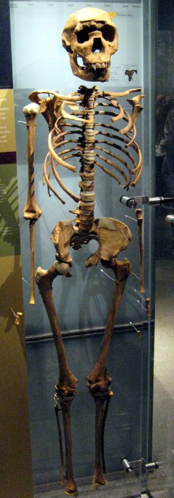
Photo courtesy of Claire Houck via Wikimedia Commons.
In modern humans, the cartilaginous growth plates at the end of each vertebrae usually fuse into bone by the late teens to mid-twenties. If the spine sustains some trauma before then (perhaps caused by an enthusiastic game of football, or carrying rocks), the intervertebral disc can herniate in such a way that the disc material slices through the bone at an angle. Sometimes, a corner of the vertebrae is snapped clean off. In other cases, the disc’s annulus can pull off a small piece of bone.
Scientists also used their detective skills to figure out what was wrong with the Nariokotome boy, who lived 1.5 million years ago. The juvenile Homo erectus had L4-L5 vertebrae that seemed off-kilter. The facet joint was unexpectedly narrow on the left side, and the bones had been remodeled to form an entirely new joint.
This originally led scientists to conclude that he had scoliosis, or spina bifida, or some other serious developmental defect. However, subsequent tests didn’t support these theories. It’s now thought that his roughed-up vertebrae were a consequence of disc herniation.
Since his facet joints were altered while the Nariokotome boy was still growing, the cause of the disc herniation was most likely traumatic (e.g., a fall from a tree, an ill-advised wrestling match) (Haeusler, Schiess and Boeni, Evidence for Juvenile Disc Herniation in a Homo Erectus Boy Skeleton 2013).
The researchers who proposed this theory noted that, “Disabling backache and recurrent sciatica might have, at least, temporarily restricted his daily activities, which indicates advanced social care and nursing in early Homo (Haeusler, Schiess and Boeni, Evidence for Juvenile Disc Herniation in a Homo Erectus Boy Skeleton 2013).”
They also noted that Homo erectus spines are narrower than those of modern-day humans, which may have made these earlier hominins more vulnerable to injury (Haeusler, Schiess and Boeni, Evidence for Juvenile Disc Herniation in a Homo Erectus Boy Skeleton 2013). Impact forces are greater when channeled through a smaller area, which is why my karate sensei told me to punch with a knuckle, rather than the flat of my fist.
The Old Man of the Pleistocene
Once we start counting years in thousands rather than millions, we see more fossils from individuals who lived longer. Their spines are notable for the number of age-related changes, as opposed to the trauma-induced injuries or developmental problems seen in earlier remains.
The oldest of these (in terms of chronological distance from us) come from Homo heidelbergensis, a species which is considered to be the last common ancestor between Neanderthals and humans. (Although this is another one of those subjects that scientists love to debate.)
One set of fossils is from a male Homo heidelbergensis who lived in Spain roughly 530,000 years ago. The remains of the SH Pelvis 1 individual consist of five lumbar vertebrae, plus the sacrum and a good deal of the pelvis. He was estimated to be over 45 years old. Which, by Pleistocene standards, made him ancient even before he died.
His lumbar vertebrae were curiously wedged forward. Some scientists thought this could be due to microfractures (which give some modern elderly people a hunched appearance). However, his bone density was just fine, so the sorts of mini-fractures caused by osteoporosis were unlikely.
His L5 vertebrae in particular was in tough shape. He had a large Schmorl’s node, a sign that the disc’s nucleus was forced into the bone in a type of herniation. His L5 vertebra had also slipped down over his S1 vertebra, though this case of spondylolisthesis was probably due to a developmental defect, rather than trauma.
The spinous processes of his L4 and L5 vertebrae had been rubbing together for some time, and showed characteristic remodeling. This condition, known as Baastrup’s disease, likely caused intense back pain.
The authors describing these maladies noted that, “Comparison with present day hunter-gatherer societies suggests this individual would likely not have participated in hunting activities. However, this theory would not imply that it was no longer capable of moving with the social group (Bonmatí, et al. 2010).”
Translation: We think his people took care of him.
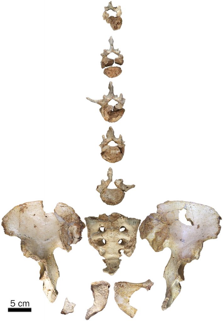
Photo from Bonmatí, et al. 2010. Copyright 2010 National Academy of Sciences.
Back Pain Among Neanderthals
Perhaps no extinct species was more committed to spinal ruin than the Neanderthals.
A Neanderthal skeleton discovered in southwestern France in the early 1900s was so arthritic that he led scientists and the public alike to think of Neanderthals as stoop-backed, barely ambulatory brutes. The actual history of The Old Man of La Chapelle was far more distinguished. He was estimated to be in his sixties when he died (give or take a decade), and bears the marks of a long, hard life.
All of his remaining facet joints show some sign of osteoarthritis, albeit to different degrees. At some levels, his facet joints are molded together in a way that suggested that the disc separating them had thinned. Three of his vertebrae have Schmorl’s nodes on both the tops and bottoms. A few of the vertebrae in his lower back had slipped slightly to the side (Haeusler, Trinkaus, et al. 2019).
The damage to the facet joints in his lumbar spine was so extensive that his lower back would have been largely immobile.
This pattern of facet joint damage and osteoarthritis is the norm for Neanderthals, although there are varying degrees of severity. An older male from La Ferrassie, France, was found with most of his vertebrae (though they were fragmentary). One scholar noted that, “All vertebrae that can be assessed show facet joint osteoarthritis (Haeusler, Spinal Pathologies in Fossil Hominins 2019).”
Another young male Neanderthal from Regourdou, France, shows facet joint osteoarthritis, as do isolated vertebrae found near Krapina, Croatia. In one case, there is evidence that these bony overgrowths might have pressed against a nerve root in the neck (Haeusler, Spinal Pathologies in Fossil Hominins 2019).
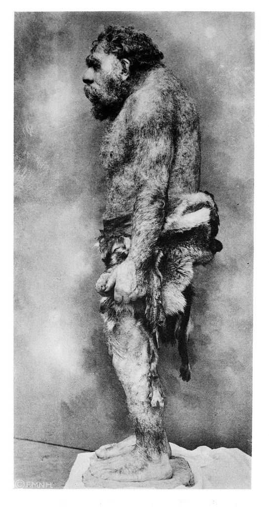
Photo courtesy of the Wellcome Collection.
If you’re interested in Neanderthals, then Shanidar Cave in Iraq is a particularly important site. The remains of eight adult and two infant Neanderthals have been recovered. Four of the adults (all males) have enough of a spine left to analyze.
Shanidar 1’s most famous injury is his withered right arm, which may be the result of an above-the-elbow amputation in his younger days. Many of his vertebrae were crushed or fragmentary, and his spine has attracted less attention. Still, scientists identified a few bony osteophytes in his neck and low back.
Shanidar 2 also shows some mild osteoarthritis, although he appeared to be in much better shape (generally speaking) than Shanidar 1. However, he also died when he was significantly younger (at 20–30 years old), so he wasn’t necessarily luckier.
Another relatively old (40–45 years old) man, Shanidar 3, has his own claim to fame – he may have been murdered. He was stabbed in the ribs, and possibly suffered a collapsed lung. In The Shanidar Neandertals, which is a generally dry book, author Erik Trinkaus gets noticeably excited when he considers the fate of Shanidar 3.
“The angle and precision of the wound make it unlikely that the injury was self-inflicted. In fact, the angle and position of the wound are close to what one would expect if a right-handed individual were to have stabbed Shanidar 3 while they were standing face to face. If this interpretation is accurate, this would be the oldest case of human interpersonal violence and the only possible one among known Neanderthals. However, the possibility that this injury resulted from interpersonal violence by no means substantiates such a claim. It is possible that Shanidar 3 injured himself accidentally, or more likely, that another member of the social group wounded him accidentally. Regardless of the circumstances surrounding the wounding, Shanidar 3 was nursed for a period of at least several weeks and was intentionally buried after his death (Trinkaus 1983).”
As for his spine, Shanidar 3 suffered the usual osteoarthritis and facet joint changes, although not to any exceptional degree.
Shanidar 4 has a badly preserved spine, but what remains shows significant osteoarthritis. He was another respectable adult, between 30–45 years old. Because his skeleton is less complete, scientists had to guess his age based on his teeth, which had most of the enamel stripped away (Trinkaus 1983).
There is one Neanderthal skeleton that got his skeletal abnormalities before he got old. Kebara 2 was estimated to be in his 30s when he died, and it appears he never developed quite right. His first lumbar vertebrae had short ribs, and his first sacral vertebrae made a halfhearted attempt to be a lumbar vertebrae. Some of his lumbar vertebrae appear to be missing the spinous process, which is probably due to a developmental defect. He had Schmorl’s node at the top of his L2 vertebrae (Haeusler, Spinal Pathologies in Fossil Hominins 2019).
Present-Day Lessons from Ancient Spines
It’s nice to think that humans evolved in some mythical Eden, where they lived peaceful and pain-free lives until they died. But perhaps that fantasy only persists because they didn’t leave us any pain pills or hospital intake forms.
Although we can’t directly ask ancient hominins to assess their pain, their bones tell us they had their own problems. They were probably no strangers to back pain. If the Nariokotome boy were alive today, I imagine he would tell me, “At least I had the good sense to die before puberty.”
So perhaps modern humans aren’t uniquely unfortunate. And at least we can work lying down.
References
Bonmatí, Alejandro, Asier Gómez-Olivencia, Juan-Luis Arsuaga, José Miguel Carretero, Ana Gracia, Ignacio Martínez, Carlos Lorenzo, José María Bérmudez de Castro, and Eudald Carbonell. 2010. “Middle Pleistocene lower back and pelvis from an aged human individual from the Sima de los Huesos site, Spain.” 107 (43): 18386-18391. doi:10.1073/pnas.1012131107.
D’Anastasio, Ruggero, Bernhard Zipfel, Jacopo Moggi-Cecchi, Roscoe Stanyon, and Luigi Capasso. 2009. “Possible Brucellosis in an Early Hominin Skeleton from Sterkfontein, South Africa.” PLoS ONE 4 (7). doi:10.1371/journal.pone.0006439.
Haeusler, Martin. 2019. “Spinal Pathologies in Fossil Hominins.” In Spinal Evolution: Morphology, Function, and Pathology of the Spine in Hominoid Evolution, by Ella Been, Asier Gómez-Olivencia and Patricia Ann Kramer, 213-245. Springer. doi:10.1007/978-3-030-19349-2_10.
Haeusler, Martin, Erik Trinkaus, Cinzia Fornai, Jonas Müller, Noémie Bonneau, Thomas Boeni, and Nakita Frater. 2019. “Morphology, pathology, and the vertebral posture of the La Chapelle-aux-Saints Neandertal.” PNAS 11: 4923-4927. doi:10.1073/pnas.1820745116.
Haeusler, Martin, Regula Schiess, and Thomas Boeni. 2013. “Evidence for Juvenile Disc Herniation in a Homo Erectus Boy Skeleton.” Spine (Lippincott Williams & Wilkins) 38 (3): E123–E128. doi:10.1097/BRS.0b013e31827cd245.
Mays, S. A. 2007. “Lysis at the Anterior Vertebral Body Margin: Evidence for Brucellar Spondylitis?” International Journal of Osteoarchaeology (Wiley InterScience) 17: 107–118. doi:10.1002/oa.903.
Meyer, Marc R., Scott A. Williams, Michael P. Smith, and Gary J. Sawyer. 2018. “Lucy’s back: Reassessment of fossils associated with the A.L. 288-1 vertebral column.” Journal of Human Evolution (Elsevier) 85: 174–180. doi:10.1016/j.jhevol.2015.05.007.
Ryan, Timothy M., and Simone Sukhdeo. 2015. “KSD-VP-1/1: Analysis of the Postcranial Skeleton Using High-Resolution Computed Tomography.” In The Postcranial Anatomy of Australopithecus afarensis, by Yohannes Haile-Selassie and Denise F. Su, 39-62. doi:10.1007/978-94-017-7429-1_4.
Sparrey, Carolyn J., Jeannie F. Bailey, Michael Safaee, Aaron J. Clark, Virginie Lafage, Frank Schwab, Justin S. Smith, and Christopher P. Ames. 2014. “Etiology of lumbar lordosis and its pathophysiology: a review of the evolution of lumbar lordosis, and the mechanics and biology of lumbar degeneration.” Journal of Neurosurgery 36 (5). doi:10.3171/2014.1.FOCUS13551.
Trinkaus, Erik. 1983. The Shanidar Neandertals. Academic Press.
Tsirikos, A I, and A K Jain. 2011. “Scheuermann’s kyphosis; current controversies.” The Bone & Joint Journal 93-B (7): 857-864. doi:10.1302/0301-620X.93B7.26129.
One thought on “Even Neanderthals Had Back Pain”